Home
/ Long Bone With Diagram : Diagram Of Long Bone Diagram Of Hand And Wrist Wrist Hand Teaching Anato Human Anatomy For Muscle Reproductive And Skeleton Sword Hero
Long Bone With Diagram : Diagram Of Long Bone Diagram Of Hand And Wrist Wrist Hand Teaching Anato Human Anatomy For Muscle Reproductive And Skeleton Sword Hero
Long Bone With Diagram : Diagram Of Long Bone Diagram Of Hand And Wrist Wrist Hand Teaching Anato Human Anatomy For Muscle Reproductive And Skeleton Sword Hero. The diaphysis is the tubular shaft that runs between the proximal and distal ends of the bone. This visually displays where a bone accepts blood vessels or where cartilage develops. The outside of the bone consists of a layer of connective tissue called the periosteum. In long bones, as you move from the outer cortical compact bone to the inner medullary cavity, the bone transitions to spongy bone. Long bones include the humerus (upper arm), radius (forearm), ulna (forearm), femur (thigh), fibula (thin bone of the lower leg), tibia (shin bone) , phalanges (digital bones in the hands and feet), metacarpals (long bones within the hand), and metatarsals (long bones.
Periosteum a dense fibrous membrane covering the surface of bones (except at their extremities) and serving as an attachment for tendons and muscles. The humeri, radii, and ulnae of the arms; Abdominal vessels anatomy 12 photos of the abdominal vessels anatomy abdominal blood vessels anatomy, abdominal circulation anatomy, abdominal vessels anatomy, abdominal wall vessel anatomy, anatomy of abdominal vessels, human anatomy, abdominal blood vessels anatomy, abdominal circulation anatomy, abdominal vessels. Add to playlist 12 playlists. Bones in spine and neck 12 photos of the bones in spine and neck bones in spine and neck, bones in the spine and neck, bone, bones in spine and neck, bones in the spine and neck
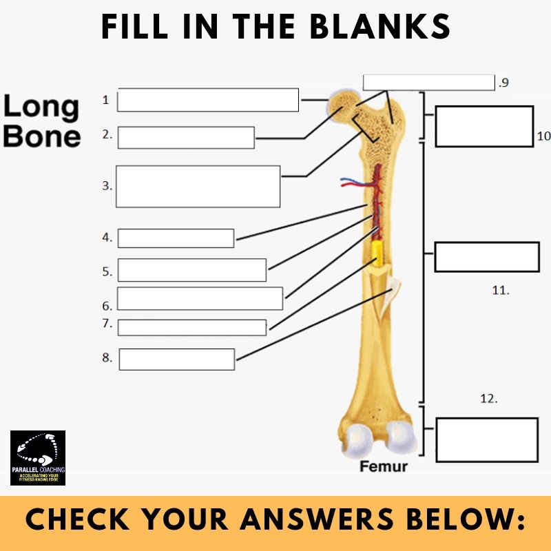
What Is The Structure Of A Long Bone L2 And L3 Anatomy Revision from parallelcoaching.co.uk #draw a well labelled diagram of a long bone #labelled diagram of a bone cell #labelled diagram of bones in the body #labelled diagram of hip bone #labelled diagram of the bone related posts of labelled diagram of long bone The long bone category includes the femora, tibiae, and fibulae of the legs; We cover the diaphysis, the epiphysis, spongy and c. You need to get 100% to score the 9 points available. A long bone has two parts: Radius and ulna bones quiz posterior markings getbodysmart at unlabeled the ulna is a bone in human forearm broader near elbow unlabeled anatomy lab photographs upper. Diagram a generic long bone and label all of the parts, including tissue types and regions. Long bones are longer than they are wide and are the major bones of the limbs.
The diaphysis is the tubular shaft that runs between the proximal and distal ends of the bone.
There is a printable worksheet available for download here so you can take the quiz with pen and paper. A hollow medullary cavity is found in the center of long bones and serves as a storage area for bone marrow. For this time we collect some pictures of long bone diagram blank, and each of them giving you some fresh ideas. This visually displays where a bone accepts blood vessels or where cartilage develops. The diaphysis and the epiphysis.the diaphysis is the tubular shaft that runs between the proximal and distal ends of the bone. This is an online quiz called long bone diagram. The diaphysis is the tubular shaft that runs between the proximal and distal ends of the bone. Diagram of of a long bone. The structure of a long bone allows for the best visualization of all of the parts of a bone (figure 1). The diaphysis and the epiphysis. Altogether, the skeleton makes up about 20 percent of a person's body weight. The end of the long bone is the epiphysis and the shaft is the diaphysis. Inside of arm muscle and bone 12 photos of the inside of arm muscle and bone , bone
It is also known as the calf bone, as it. Compact bone is the hard material that makes up the shaft of long bones and the outside surfaces compact bone consists of cylindrical units called osteons. Long bones are longer than they are wide and are the major bones of the limbs. A long bone has a shaft and 2 ends. Next to the tibia is the fibula, the thinner, weaker bone of the lower leg.
Anatomy Physiology Midterm Review Study The Long Bone Image And Identify The Parts Of The Bone Diagram Quizlet from o.quizlet.com When showing this long bone diagram blank, i can guarantee to rock your world!. Metacarpals and metatarsals of the hands and feet, the phalanges of the fingers and toes, and the clavicles or collar bones. The interior part of the long bone is the medullary cavity with the inner core of the bone cavity being composed of marrow. Next to the tibia is the fibula, the thinner, weaker bone of the lower leg. Used figure 6.2 in book. The human skeletal system consists of all of the bones, cartilage, tendons, and ligaments in the body. #draw a well labelled diagram of a long bone #labelled diagram of a bone cell #labelled diagram of bones in the body #labelled diagram of hip bone #labelled diagram of the bone related posts of labelled diagram of long bone Radius and ulna bones quiz posterior markings getbodysmart at unlabeled the ulna is a bone in human forearm broader near elbow unlabeled anatomy lab photographs upper.
The other primary skeletal component of height are the vertebrae and skull.
Bone growth diagrams show the progression of development of the bone over a period of time. Periosteum a dense fibrous membrane covering the surface of bones (except at their extremities) and serving as an attachment for tendons and muscles. Long bones are one of the five bone types that are classified by shape. Learn long bone diagram with free interactive flashcards. Related posts of diagram of of a long bone inside of arm muscle and bone. A typical long bone shows the gross anatomical characteristics of bone. The other primary skeletal component of height are the vertebrae and skull. Radius and ulna bones quiz posterior markings getbodysmart at unlabeled the ulna is a bone in human forearm broader near elbow unlabeled anatomy lab photographs upper. Inside of arm muscle and bone 12 photos of the inside of arm muscle and bone , bone A long bone has two parts: Examples of long bones include the femur, tibia, fibula, metatarsals, and phalanges. Parts of long bone (applies to other bones too). We cover the diaphysis, the epiphysis, spongy and c.
You need to get 100% to score the 9 points available. The structure of a long bone allows for the best visualization of all of the parts of a bone (figure 6.7). The other primary skeletal component of height are the vertebrae and skull. The long bone category includes the femora, tibiae, and fibulae of the legs; Bone growth diagrams show the progression of development of the bone over a period of time.
Long Bone Internal Structure Human Anatomy Body Human Bones Anatomy Human Body Systems Human Body Anatomy from i.pinimg.com The outside of the flat bone consists of a layer of connective tissue called the periosteum. A long bone is a bone that has greater length than width. The diaphysis and the epiphysis. In this video we discuss the parts of a long bone and some of the functions of each of those bone parts. The outside of the bone consists of a layer of connective tissue called the periosteum. Online quiz to learn long bone parts quiz; Long bones have a thick outside layer of compact bone and an inner medullary cavity containing bone marrow. Related posts of long bone anatomy diagram bones in spine and neck.
Used figure 6.2 in book.
Altogether, the skeleton makes up about 20 percent of a person's body weight. Compact bone is the hard material that makes up the shaft of long bones and the outside surfaces compact bone consists of cylindrical units called osteons. Structure of the long bone with pictures learn with flashcards, games, and more — for free. A hollow medullary cavity is found in the center of long bones and serves as a storage area for bone marrow. Related posts of diagram of of a long bone inside of arm muscle and bone. The other primary skeletal component of height are the vertebrae and skull. The structure of a long bone allows for the best visualization of all of the parts of a bone ().a long bone has two parts: The humeri, radii, and ulnae of the arms; Online quiz to learn long bone parts quiz; Broken left elbow bone 12 photos of the broken left elbow bone broken elbow bone spur, broken elbow bone symptoms, bone, broken elbow bone spur, broken elbow bone symptoms. Long bones have a thick outside layer of compact bone and an inner medullary cavity containing bone marrow. This is an online quiz called long bone anatomy. The end of the long bone is the epiphysis and the shaft is the diaphysis.
Share :
Post a Comment
for "Long Bone With Diagram : Diagram Of Long Bone Diagram Of Hand And Wrist Wrist Hand Teaching Anato Human Anatomy For Muscle Reproductive And Skeleton Sword Hero"


Post a Comment for "Long Bone With Diagram : Diagram Of Long Bone Diagram Of Hand And Wrist Wrist Hand Teaching Anato Human Anatomy For Muscle Reproductive And Skeleton Sword Hero"